Excitation and Emission
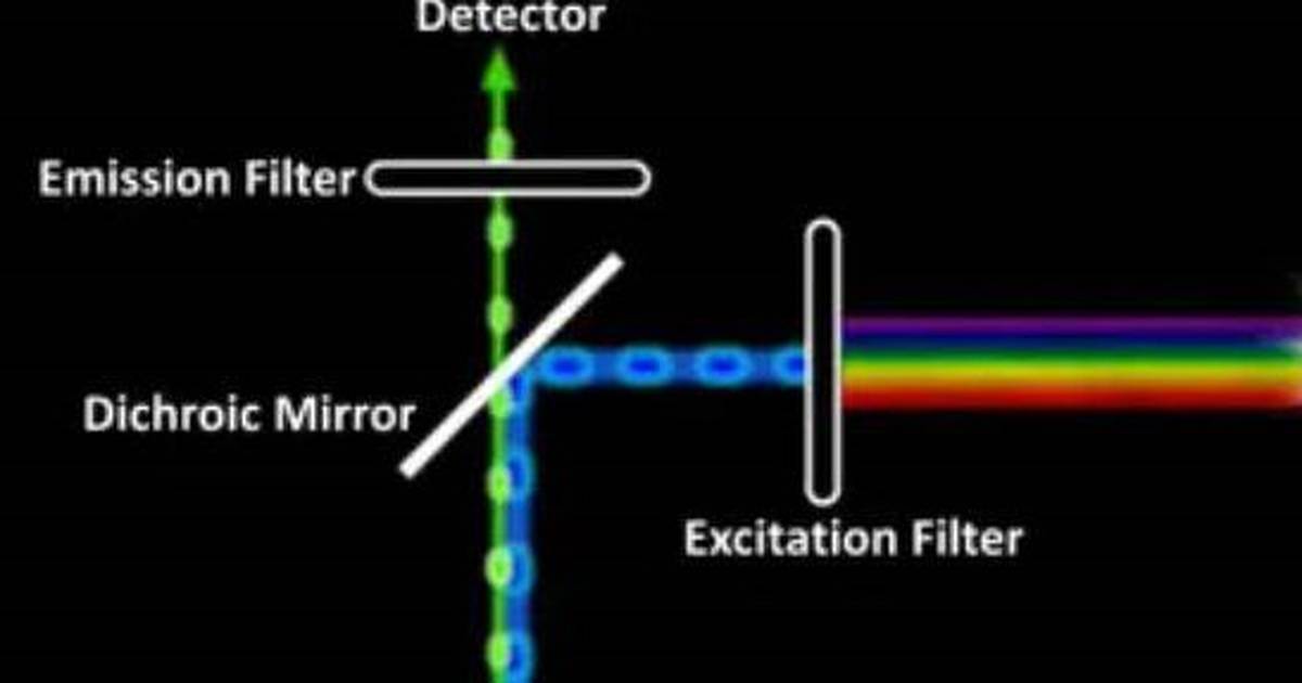
In fluorescence microscopy, a specimen is typically labeled with a fluorescent molecule, or fluorophore. When illuminated with light of a specific wavelength, the fluorophore can be excited and will subsequently emit light of a longer wavelength. This emitted light is detected by a CCD, and the image is captured. This animation shows a simple animated diagram of a fluorescence microscope.
Topics
- fluorescence
- microscopy
- electromagnetic spectrum
- CCD
- fluorophore
- apparatus
- emission
- excitation emission
- detector
- filter
- objective
- specimen
- dichroic mirror